Nov 3, 2023The knee joint is a hinge type synovial joint, which mainly allows for flexion and extension (and a small degree of medial and lateral rotation). It is formed by articulations between the patella, femur and tibia. In this article, we shall examine the anatomy of the knee joint – its articulating surfaces, ligaments and neurovascular supply.
Knee Osteotomy Surgery Procedure | Arthritis-health
Human Anatomy Laboratory Manual 2021 7: Joints 7.2: Knee Joint

Source Image: pinterest.com
Download Image
Policy Function What does the knee joint do? Your knees have several important jobs, including: Moving your legs. Supporting your body when you stand and move. Stabilizing you and helping keep your balance. Anatomy Where is the knee joint located? The knee is the joint in the middle of your leg.
Source Image: teachmeanatomy.info
Download Image
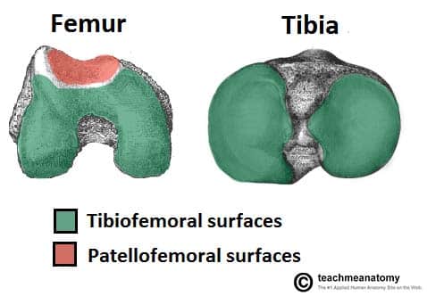
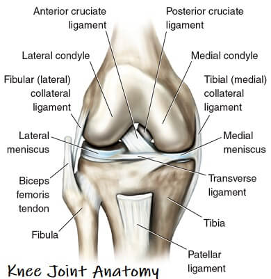
Knee joint anatomy Nov 5, 2023The patella is a sesamoid bone—the largest in the body—occupying the anterior part of the knee. It is the distal attachment point of the quadriceps tendon. The patella also protects the anterior articular surface of the distal femur. The fibula is not part of the knee joint. The surfaces articulating at the knee are the following:

Source Image: 123rf.com
Download Image
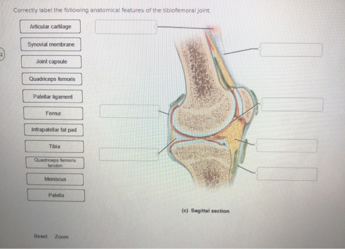
Correctly Label The Following Anatomical Features Of The Knee Joint.
Nov 5, 2023The patella is a sesamoid bone—the largest in the body—occupying the anterior part of the knee. It is the distal attachment point of the quadriceps tendon. The patella also protects the anterior articular surface of the distal femur. The fibula is not part of the knee joint. The surfaces articulating at the knee are the following: Dec 27, 2023The main features of the knee anatomy include bones, cartilages, ligaments, tendons and muscles. In the knee joint, the femur articulates with the tibia and the patella. The knee joint is a synovial joint this means it contains a fluid that lubricates it. This fluid is known as the synovial fluid.
Anatomy. Knee Joint Cross Section Showing The Major Parts Which Made The Knee Joint For Basic Medical Education Also For Clinics Royalty Free SVG, Cliparts, Vectors, and Stock Illustration. Image 64101407.
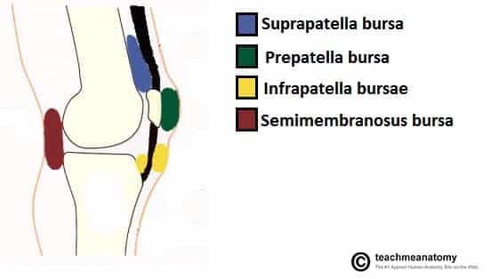
Correctly label the following anatomical features of the knee joint Propatollar bursa Bursa deep to gastrocnemius muscle Suprapatellar bursa Moniscus Infrapatellar bursa This problem has been solved! You’ll get a detailed solution from a subject matter expert that helps you learn core concepts. See Answer The Knee Joint – Articulations – Movements – Injuries – TeachMeAnatomy
Source Image: teachmeanatomy.info
Download Image
Solved Correctly label the following anatomical features of | Chegg.com Correctly label the following anatomical features of the knee joint Propatollar bursa Bursa deep to gastrocnemius muscle Suprapatellar bursa Moniscus Infrapatellar bursa This problem has been solved! You’ll get a detailed solution from a subject matter expert that helps you learn core concepts. See Answer
Source Image: chegg.com
Download Image
Knee Osteotomy Surgery Procedure | Arthritis-health Nov 3, 2023The knee joint is a hinge type synovial joint, which mainly allows for flexion and extension (and a small degree of medial and lateral rotation). It is formed by articulations between the patella, femur and tibia. In this article, we shall examine the anatomy of the knee joint – its articulating surfaces, ligaments and neurovascular supply.
Source Image: arthritis-health.com
Download Image
Knee joint anatomy Policy Function What does the knee joint do? Your knees have several important jobs, including: Moving your legs. Supporting your body when you stand and move. Stabilizing you and helping keep your balance. Anatomy Where is the knee joint located? The knee is the joint in the middle of your leg.

Source Image: pinterest.com
Download Image
Knee Joint Anatomy: Structure, Function & Injuries – Knee Pain Exp Chapter Outline Osteology Distal Femur Proximal Tibia Proximal Fibula Patella Arthrology General Features Normal Alignment Supporting Structures Kinematics Muscle and Joint Interaction Innervation of the Muscles of the Knee Muscles of the Knee Internal and External Rotators of the Knee Summary Study Questions Additional Readings Objectives
Source Image: knee-pain-explained.com
Download Image
Solved Correctly label the following features of the knee | Chegg.com Nov 5, 2023The patella is a sesamoid bone—the largest in the body—occupying the anterior part of the knee. It is the distal attachment point of the quadriceps tendon. The patella also protects the anterior articular surface of the distal femur. The fibula is not part of the knee joint. The surfaces articulating at the knee are the following:

Source Image: chegg.com
Download Image
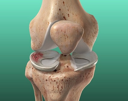
Meniscus Injury: Torn Knee Cartilage – Causes & Treatment Dec 27, 2023The main features of the knee anatomy include bones, cartilages, ligaments, tendons and muscles. In the knee joint, the femur articulates with the tibia and the patella. The knee joint is a synovial joint this means it contains a fluid that lubricates it. This fluid is known as the synovial fluid.
Source Image: knee-pain-explained.com
Download Image
Solved Correctly label the following anatomical features of | Chegg.com
Meniscus Injury: Torn Knee Cartilage – Causes & Treatment Human Anatomy Laboratory Manual 2021 7: Joints 7.2: Knee Joint
Knee joint anatomy Solved Correctly label the following features of the knee | Chegg.com Chapter Outline Osteology Distal Femur Proximal Tibia Proximal Fibula Patella Arthrology General Features Normal Alignment Supporting Structures Kinematics Muscle and Joint Interaction Innervation of the Muscles of the Knee Muscles of the Knee Internal and External Rotators of the Knee Summary Study Questions Additional Readings Objectives